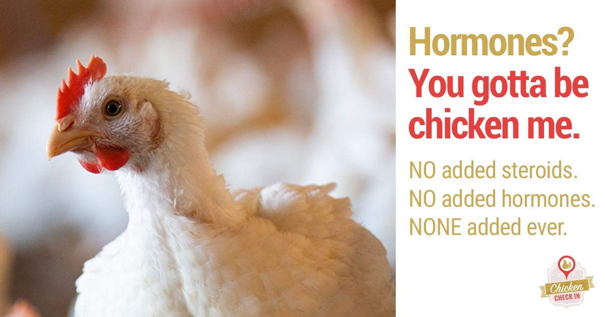
People all over the world love chicken because it can be used in many different ways, is cheap, and is a good source of lean protein. However, for years, rumors have been going around that hormones are used to make chickens grow bigger and faster. This has made many people wonder if there are hormones in chicken meat.
The short answer is no The use of added hormones in poultry production has been illegal in the United States since the 1950s. However, confusion persists due to misunderstandings about poultry farming practices and naturally occurring hormones in chickens
This article will clarify why hormones are not used in chicken production, explain the real reasons behind rapid chicken growth, and discuss naturally occurring hormone levels in poultry.
Why Hormones Are Not Used in Chicken Production
-
The U. S. Between 1950 and 1970, the U.S. Department of Agriculture (USDA) banned the use of hormones to make chickens grow faster. Regulations strictly prohibit the administration of hormones in poultry farming.
-
The Food and Drug Administration (FDA) does not allow the use of hormones to raise chickens, turkeys, or other birds. Labels on chicken that say “no hormones added” are not allowed unless they are followed by a statement that says federal laws do not allow the use of hormones.
-
Unlike cattle farming, hormone use provides no benefits for chicken production. Chickens reach their growth potential naturally through selective breeding and optimized nutrition.
-
Administering hormones to chickens would require impractical multiple daily injections per bird. Modern production systems make this infeasible.
-
Using hormones could also cause health issues like lameness and mortality. Hormones would force unnatural, dangerous growth beyond chickens’ physiological limits.
The Real Reasons Behind Rapid Chicken Growth
Instead of hormones, three main factors enable the fast growth seen in commercial chickens:
Selective Breeding
- Over decades, chicken producers selectively bred birds to achieve rapid growth and efficient feed conversion. Short generation intervals enabled rapid selection of desired traits.
Optimized Nutrition
- Precisely formulated feeds provide chickens exactly the right amounts of energy, protein, vitamins, and minerals for maximum performance.
Ideal Growing Environments
- Barns offer ideal temperature, ventilation, lighting, and space for chickens to thrive. This environment complements birds’ genetics and diet.
Naturally Occurring vs. Added Hormones
-
Chickens, like all animals, make small amounts of hormones like estrogen, progesterone, and testosterone on their own. These are essential for normal functioning.
-
However, regulations prohibit supplementing chicken feed with additional hormones beyond their natural levels.
-
In fact, each day the human body secretes vastly larger amounts of hormones than are ingested from all food sources combined.
-
Any hormones consumed through eating chicken or other meats are efficiently deactivated and metabolized by the body, limiting their physiological impact.
Persistent Confusion and Misconceptions
Despite the facts, misunderstandings around hormone use in poultry farming have persisted due to:
-
Beef cattle: Growth hormones are commonly used in beef cattle production, which is legal and regulated for that industry. This causes confusion between cattle and chicken production.
-
Lack of consumer awareness: Many consumers simply do not know that federal policies prohibit chicken hormone use and wrongly assume it is a common practice.
-
Natural vs. added: The difference between natural and added hormones is often poorly understood.
-
Rapid growth: Some consumers see the rapid growth achieved through breeding, nutrition and housing and wrongly assume it must be due to hormones.
The Bottom Line: No Hormones in Chicken
While chickens contain small, natural levels of hormones, the use of additional hormones in poultry farming was banned in the 1950s and remains illegal. Rapid chicken growth stems from selective breeding, optimized nutrition, and ideal housing – not hormones. Consumers can rest assured that chicken sold in the U.S. contains no added hormones.
Prolactin and the Immune System
PRL is a 23-kDa polypeptide hormone released by anterior pituitary cells. It is very similar in many types of poultry (Kansaku et al. , 2008; Wang et al. , 2009). PRL also exists in thymus, spleen, lymphocytes, and epithelial cells (Bolefeysot et al. , 1998; Wilkanowska et al. , 2014). The chicken PRL is mainly related to nesting, hatching, egg production, broodiness (Jiang et al. , 2005; Cui et al. , 2006; Rashidi et al. , 2012; Li et al. , 2013; Wilkanowska et al. , 2014).
The levels of PRL in plasma is different in various growth periods of chicken (Jiang et al. , 2005; Wilkanowska et al. , 2014). Even before and after ovulation, there are significant changes (*P < 0. 05) in the PRL concentrations (Scanes et al. , 1977). Meanwhile, PRL levels alters in different chicken breeds (Mo et al. , 2022). When chickens are infected with pathogens, such as Histomonas meleagridis (Chadwick et al. , 1980), E. tenella (Chadwick et al. , 1985), and ALV-J (Mo et al. , 2021), the plasma PRL levels have changed compared with the uninfected chickens. These results indicate that PRL levels is not only affected by individual own growth cycle but also controlled by external factors.
PRL is a cytokine that stimulates cellular and humoral immunity at the same time. At the 14th days post-infection (dpi) infected with Trypanosoma cruzi (T. When rats are infected with T. cruzi, PRL treatment raises the number of natural killer (NK) cells and B lymphocytes in their spleen compared to rats that were infected without PRL treatment and rats that were not treated (Filipin et al. , 2019). Studies have shown that PRL controls immune cells when parasites infect them, boosts the immune system of the host, and lowers the harmful effects of pathogens (Filipin et al. , 2011, 2019; Río-Araiza et al. , 2018a,b). PRL promotes lymphocyte mitogenesis isolated from thymus and spleen of White Leghorn chickens in a dose-dependent manner (SkwarłoSońta, 1990). Different doses of ovine PRL were used to treat chicken bursa of fabricius cells, and all doses of PRL increase the mitotic activity of cells, and the lowest dosage of PRL is most effective (Bhat et al. , 1983). Also, PRL changed the difference between chemotactic factor and leukocyte migration of fetal membranes in a way that was specific to each tissue (Núñez-Sánchez et al. , 2021). Our earlier research showed that adding PRL to DF-1 cells before they are infected with ALV-J has the strongest antiviral effects in vitro, no matter what dose was used (Mo et al. , 2021). Moreover, PRL can reduce the expression of pro-inflammatory cytokine-encoding TNTα, IL-1β, and IL-6 genes, and increase the expression of the interferon-stimulated genes of oligoadenylate synthetase-like (OSAL) and vasoactive intestinal peptide (VIP) in the spleens of ALV-J-infected chicks (Mo et al. , 2021). Interestingly, hormone deficiency can alter the host immune system and reduce specific antibodies against pathogens (Quintanar-Stephano et al. , 2015; Hernández-Cervantes et al. , 2018; Río-Araiza et al. , 2018a,b). Different PRL levels in different breeds may also explain why some breeds are more or less likely to get viruses (Mo et al. , 2022). PRL plays an important role in regulating and maintaining the host immune response, and is an indispensable part of the host immune system.
During the lactation, PRL can increase the number of immune cells in mammary secretions and enhance the chemotaxis effect on T cells, B cells, monocytes, and macrophages (Dill and Walker, 2017). In addition, PRL can reduce the activation threshold of B cell receptors and induce CD40 expression in B cells (Correale et al., 2014). During the first hour of bacterial infection, PRL increases the expression of TLRs and MyD88, while the expression of IL-1β is continuously increased (Peña et al., 2016). Pituitary cells can directly recognize the fungal cell wall glucans to promote the expression of TLR4 and CD14 (Breuel et al., 2004). During T. cruzi infection, PRL increase the NK cells levels in treated infected animals compared to the untreated group (Filipin et al., 2019). Furthermore, PRL could reduce lipopolysaccharide (LPS)-induced inflammatory cytokines (TNF-α, IL-1β, and IL-6) via inhibiting NFκB phosphorylation and TLR4 expression (Olmos-Ortiz et al., 2019). However, the inhibitory effect of PRL is selective (Flores-Espinosa et al., 2020). PRL also stimulates the internalization of staphylococcus aureus on bovine mammary epithelial cells, and up-regulates the mRNA expression of TNF-α, IL-1β, and inducible nitric oxide synthase (Gutiérrez-Barroso et al., 2008). PRL constrains tumor-promoting liver inflammation by inhibiting MAP3K-dependent activation of c-Myc at the level of the “trafasome” (comprised of IRAK1, TRAF6, and MAP3K proteins) (Hartwell et al., 2014). However, the macrophages exposed with PRL secrete more inflammatory factors and reactive oxygen species, thereby aggravates the inflammatory response (Majumder et al., 2002; Sodhi and Tripathi, 2008; Tang et al., 2016). These results suggest that PRL not only affects the differentiation, regulation, and responsiveness of immune cells, but also the secretion of different immune messengers, such as chemokines, interleukin, and interferon, and increases layers of complexity to the interactive molecular and cellular events that occur in inflammatory and virus infectious diseases (Recalde et al., 2018).
PRL gene transcription is activated by the cyclic adenosine monophosphate (cAMP)-induced factor(s) and pituitary-specific transcription factor 1 (Pit-1) (Ohkubo et al., 2000). PRL secretion in poultry is under stimulatory control exerted by the hypothalamus (AlKahtane et al., 2003; Wilkanowska et al., 2014). Therefore, PRL secretion is predominantly regulated by the VIP, dopamine (DA), and serotonine (5-HT) (Macnamee et al., 1986; Kagya-Agye et al., 2012). VIP is a pleiotropic neuropeptide released by neurons and immune cells, which is widely distributed and expressed in all tissues and organs, and plays an important role in inflammation and autoimmune suppression diseases (Delgado and Ganea, 2011; Ganea et al., 2014). The viral infection affects the expression of VIP in immune organs. After chicks were infected with ALV-J, the VIP mRNA expression in the spleen was correlated with the levels of PRL in plasma (Mo et al., 2021).
Many cytokines are produced in the brain, hypothalamus, or pituitary gland, such as interferon (IFN)-α, IFN-γ, IL-1, IL-2, IL-6, IL-18 and TNF-α (Breder et al., 1988; Petrovsky, 2001; Silverman et al., 2005; Borghetti et al., 2009). These factors can stimulate or inhibit hormone secretion in the central nervous system (Rothwell and Hopkins, 1995; Steinmann, 2004; Borghetti et al., 2009). Furthermore, chemokines and their receptors also play regulatory roles in the neuroendocrine system (Callewaere et al., 2007). IL-1, IL-2, and IL-6 can stimulate PRL secretion, while endothelin-3 and IFN-γ play an inhibitory influence (Chikanza, 1999; Borba and Shoenfeld, 2019). The fungal cell wall glucans also promote the secretion of PRL (Breuel et al., 2004). LPS might promote the secretion of PRL by anterior pituitary cells through the mediation of IL-6 (Tomaszewska-Zaremba et al., 2018). However, cytokines IL-1β, IL-2, and IL-4 reduce PRL expression in T lymphocytes (Gerlo et al., 2005). ALV-J causes chicken monocyte cells death and accompanies with increased IL-1β and IL-18 expression (Dai et al., 2017). Moreover, the PRL, IL-1β, and IL-6 expression is also higher than that of uninfected individuals (Mo et al., 2021). Apparently, cytokines act as a two-way communication between the immune and endocrine systems. There might be a possible that the elevated expression of pro-inflammatory cytokines induced by virus infection may affect the PRL expression in vitro. The expression of proinflammatory factors (mainly IL-1, IL-6, and TNF-α) is up-regulated during the acute phase reaction (APR) in infection. These pro-inflammatory cytokines stimulate the neuroendocrine system, thereby enhance innate immunity, induce metabolic changes, and control inflammation to restore body balance (Berczi et al., 1998). In fact, cytokines produced during infection indeed play a regulatory role in coordinating the endocrine and immune systems.
PRL is not only a pituitary hormone that plays a major role in reproduction, but also acts as a cytokine in the immune response. PRL can influence the local environment of immune organs and contribute to the maturation and function of immune cells. The presence of PRL significantly improves the proliferation of immune cells (Fojtíková et al., 2010). However, the effects of PRL on the immune system are contradictory. It inhibits lymphocyte proliferation at high concentrations and enhances proliferation at low concentrations (Matera et al., 1992; Nela et al., 2014; Suarez et al., 2015). In CD4+ T cells, low-dose of PRL induces T-bet expression, but high-dose of PRL exhibits an opposite effect (Tomio et al., 2008). It means that different doses of PRL will have different effects on downstream responses (Tomio et al., 2008; Zhang et al., 2019). Therefore, it is possible that PRL regulates host immunity over a very narrow range of PRL concentrations. The correlation between PRL and autoimmune diseases such as systemic lupus erythematosus, rheumatoid arthritis, systemic sclerosis, and multiple sclerosis, has been confirmed, in which the excessive of PRL expression aggravates the symptoms of autoimmune diseases (Vera-Lastra and Jara, 2002; Borba et al., 2018; Borba and Shoenfeld, 2019). Thus, this situation may also exist in poultry. The high expression of PRL may enhance the immune response of birds, but the continuous high expression may lead to the occurrence of autoimmune diseases in poultry.
PRL itself cannot initiate the immune response, and mainly maintains the balance of immune response in the body (Fojtíková et al., 2010). PRL can triggers different reaction pathways (Gala, 1991; Bolefeysot et al., 1998). Activation of these pathways results in endpoints such as differentiation, proliferation, survival, secretion, and immune (Bolefeysot et al., 1998; Kansaku et al., 2008). This supports the hypothesis that when pathogens invade and attack host cells, cell damage or death leads to increase the expression of pro-inflammatory cytokines such as interleukins, chemokines, interferons, and tumor necrosis factors, and then the pro-inflammatory cytokines increase or inhibit PRL expression/secretion by regulating hypothalamus or immune cells. The combination of PRL and PRLR activates JAK/STAT, MAPK, and PI3K signaling pathways, thus regulating the individual immune response (Figure 1). However, there are still many mechanisms in this process that remains unknown, such as the mechanism by which pro-inflammatory factors promote or inhibit the secretion of PRL? When facing different pathogens, which signaling pathway is activated by PRL?
Schematic representation of PRL signaling pathway after virus infection. After infecting host cells, the virus causes immune cells death and increase the expression of inflammatory factors, such as chemokines, interleukin, and interferon. Inflammatory factors stimulate the pituitary gland to increase PRL secretion. In addition, cells can increase PRL secretion by autocrine or paracrine pathways. Then PRL binds to the PRLR receptor on the cell membrane, and activates JAK/STAT, MAPK, and PI3K signaling pathways, thus regulating the individual immune response.
Growth Hormone and the Immune System
Growth hormone (GH) is a 22 kDa single chain polypeptide hormone synthesized and secreted by eosinophils in the anterior pituitary. The GH gene sequences in different poultry breeds have high homology, but low homology compared with mammals (Lamb et al., 1988; Tanaka et al., 1992). Except for pituitary gland, GH also expresses in various tissues and cells, such as spleen, thymus, ovary, kidney, liver, and lymphocytes (Gala, 1991; Liu et al., 2001). These results suggest that in addition to the endocrine mode of action, there is an autocrine or paracrine mode in tissues and cells. GH accelerates muscle and bone growth, protein synthesis and fat decomposition in animals. Moreover, GH regulates gender differentiation, sexual maturity, pregnancy, lactation, and reproduction (Stephen et al., 2001; Rotwein and Chia, 2010).
The secretion of GH is directly regulated by the hormones of hypothalamus, such as growth hormone releasing hormone (GHRH) and somatostatin (SRIF). Ghrelin, a GH-releasing peptide, is a powerful endogenous GH-secretin (Peino et al., 2000; Seoane et al., 2000). Ghrelin can induce GH secretion by activating the growth hormone secretagogue receptor in the hypothalamus and pituitary gland (Reichenbach et al., 2012). IL-1 has a direct effect on the hypothalamic-pituitary-adrenal (HPA) axis at both the hypothalamic and pituitary level, leading to increase in plasma concentrations of GH, luteinizing hormone (LH), and follicle-stimulating hormone (FSH) (Sapolsky et al., 1987; Beach et al., 1989). TNF-α can induce GH secretion in APR (Berczi et al., 1998; Borghetti et al., 2009). GH can promote the IL-6 expression in the thymus, while IL-6 can stimulate hypothalamus and induce the pituitary to secrete GH (Tsigos et al., 1997). As IL-6 receptors are distributed in the brain, pituitary gland, and adrenal gland (Hopkins, 2007), GH may respond rapidly to innate immune responses to pathogens by increasing levels of related cytokines. Cytokines act as a molecular signal transduction between GH secretion and the immune system. Other factors, such as breed, age, and other conditions (temperature, light, and nutrition level) also affect the GH secretion (Harvey et al., 1978; Castaño et al., 2005).
Like the expression of PRL, viral infection also affects the expression of GH in plasma. Five-week-old piglets immunized with porcine reproductive and respiratory syndrome virus (PRRSV) exhibit the increased levels of GH, pro-inflammatory and pro-immune cytokines in the plasma compared with the control group (Borghetti et al., 2011). The plasma levels of GH in 4-week-old piglets infected with highly pathogenic PRRSV is much higher than that of blank control group and conventional strain group at 7–21 dpi (Saleri et al., 2019). After infection with human immunodeficiency virus (HIV), the spontaneous GH secretion of patient and the GH response to the stimulation are attenuated (Rochira and Guaraldi, 2017). In antiretroviral therapy (cART), the combined use of GH can reduce the bodys immune activation, improve CD4+ T lymphocyte count and HIV-1-specific T cell response (Herasimtschuk et al., 2014). The mice with generalized ablation of GHRH gene (Ghrh -/-) are highly susceptible to Streptococcus pneumoniae, but have normal thymus and T-cell development (Bodart et al., 2018). After ALV-J infection, the plasma of GH levels is higher than that of uninfected chickens (Mo et al., 2022). The interaction between GH and the immune system has been demonstrated in a variety of domestic animals (Borghetti et al., 2009). These results indicate that GH also plays an important role in the immune system of chickens. GH can regulate the immune response of the body. Treatment of autoimmune thyroiditis with recombinant chicken GH increases the proportion of CD4+ and CD8+ in thymocytes (Marsh et al., 1992). GH affects the growth and development of thymus and the maturation of lymphocytes in thymus, but has no effect on bursa of fabricius (Johnson et al., 1993). Continuous injection of GH into chicks for a week, the blastogenic response of lymphocytes to concanavalin-A or LPS mitotic stimulation is significantly increased (Haddad and Mashaly, 1992). Recombinant bovine GH (rbGH) can be used as an immunomodulator against E. tenella infection in chickens, but has no effect on E. cervulina or E. maxima (Allen et al., 1997). However, the rbGH regulates the weight growth of chicks depending on the dosage (Allen and Danforth, 1997). Injection of recombinant bovine somatotropin into peripartum dairy cows improves the innate immune response and the IgG concentration of cows, but has limited influences on metabolism (Silva et al., 2015). In MDV infected cells, GH co-expresses with the proteins encoded by MDV serotype 1 (virulent) strains SORF2 gene, and the polymorphism of GH gene is associated with the number of tissues with tumors in commercial White Leghorn chickens (Liu et al., 2001). In chicken bursa cells, the anti-apoptotic effect of GH is mediated by PI3K/Akt pathway (Luna-Acosta et al., 2015). There are evidences showing that GH is involved in the development, differentiation, and regulation of the immune response, and can be used as an immunomodulator.
Generally, GH exerts its function by regulating downstream gene expression in target cell that indirectly controls the expression of downstream genes. A previous study has shown that GH regulates cell growth, proliferation, and tissue regeneration by affecting the expression of a series of genes related to cells growth (Rotwein and Chia, 2010). The mRNA expression of 8 genes, including gp130, STAT4, and MAPK p38, is upregulated after GH injection in pituitary deficient rats (Thompson et al., 2000). GH may protect lymphocytes from glucocorticoid-induced apoptosis by inhibiting the activation of NFκB signaling pathway, or activating NFκB signaling pathway to participate in autoimmune diseases (Jeay et al., 2001, 2002). Like PRL, GH also activates ERK, MAPK and PI3K signaling pathways (Halevy et al., 1996; OConnor, 2003; Reindl et al., 2011).
GH has two distinct effects: a direct effect is GH binding to its receptor on target cells and an indirect effect is mediated primarily by insulin-like growth factor 1 (IGF1). The growth promoting effect of GH is mediated to a large extent by IGF1. GH/IGF1 has a prominent regulatory role in the immune response to infection, and mainly influences humoral and cellular functions (Heemskerk et al., 1999). IGF1 can regulate adaptive immunity, mainly by stimulating lymphopoiesis and increasing the responses to antigen-mediated activation. In mammals, IGF1 is an important cytokine that stimulates the hosts immune response, including antibody production, lymphocyte proliferation, phagocytosis, and natural killer cell activity (Auernhammer and Strasburger, 1995; Jeay et al., 2002; Yada, 2007). GH combines with GHR to activate the synthesis of IGF1 by the phosphorylation of JAK2/STAT5 (Hu et al., 2021). It has been proven that GH, directly or indirectly through the IGF1, influences both cellular immunity and humoral immunity (Hakuno and Takahashi, 2018; Szalecki et al., 2018). These results suggest that some immune effects of GH may be regulated by IGF1. The GH-GHR-IGF1 axis has been recognized to play essential roles in somatic growth, including cell proliferation, differentiation, division, and survival. On the other hand, the GH-GHR-IGF1 axis also plays an important role in immune function, with regulatory mechanisms of unexpected complexity and versatility (Figure 2) (Hu et al., 2021).
Schematic representation of GH signaling pathway after virus infection. The interactions between GH and cytokines are ambiguous and contradictory but the ghrelin can induce GH secretion. After virus infection, the host cells increase the expression of ghrelin. Ghrelin stimulates the pituitary gland to increase GH secretion. Meanwhile, cells can also increase GH secretion by autocrine or paracrine pathways. Then GH binds to the GHR receptor on the cell membrane, and activates JAK/STAT, MAPK and PI3K signaling pathways, thus regulating the individual immune response. Moreover, GH combined with GHR to activate the IGF1by inducing the JAK2/STAT5 in response to immune response.
Meanwhile, GH also affects the progress of autoimmune disease. GH slows the progression of diabetes by reducing apoptosis and/or increasing the proliferation rate of insulin-producing β cells (Villares et al., 2013). When individual against bacterial infection, GH can enhance the phagocytic activity, killing and elimination of bacteria via increasing opsonic activity (Saito et al., 1996). However, whether there is a similar response when the chickens encounter a virus infection remains unknown.
Immune cells can secrete numerous bioactive cytokines which affect neuroendocrine processes, but the activity of immune system is also modulated by cytokines (Pagani et al., 2005). The interactions between GH and cytokines are ambiguous and contradictory in the available literature (Szalecki et al., 2018). During viral infection, the mechanism of feedback to the hypothalamus remains unclear. Studies have shown that the expression of ghrelin in plasma and bursa of fabricius is upregulated in virus-infected individuals (Yu et al., 2019). Most importantly, ghrelin can induce GH secretion (Reichenbach et al., 2012). But not all virus increases the expression of ghrelin. We support the hypothesis that the combination of GH and GHR might activate JAK/STAT, MAPK or PI3K signaling pathways to regulate the host immune response after viral infection (Figure 2).
The Truth About Hormones in Chicken
FAQ
Do they still put hormones in chicken?
No, hormones are not used in chicken production in the United States. The use of hormones and steroids in poultry production has been banned since the 1950s by the USDA.
Do chickens have a lot of hormones?
Often called “broiler chickens,” people think that chickens raised for food are pumped full of steroids or artificial growth hormones to make them fatter. While the vast majority of Americans believe this to be the case, it’s just not true.
Do hormones in chicken affect humans?
Although the natural presence of estradiol, progesterone, and testosterone in the human body and their role in physiological function regulation are known, consumption of meat and meat products derived from livestock treated with the mentioned hormones can cause exposure of consumers to various levels of the residues, .
What meats have hormones in them?
The takeaway here is that there will always be some level of naturally occurring hormones present in meat, whether it’s beef, pork or poultry. But when it comes to beef, look for the label of “no added hormones” and rest assured that all poultry and pork will always be hormone free, label or not.
Are there hormones in chickens?
Myth Busted: The Real Reason Behind Poultry Growth – No Hormones, Just Science! In poultry production, there’s often confusion about the use of hormones, steroids, and questions about the genetic modification of chickens, due to some of larger piece sizes consumers sometimes see, when shopping for chicken products. (No!) Added Hormones and Steroids.
Are chickens hormone-free?
Dr. Macklin: No chickens raised in the U. S. or internationally, are given hormones. Feeding chickens additional hormones has been illegal in the U. S. since the 1950s, and it’s just not a cost-effective way to raise chickens. Why do some companies label chicken as hormone-free?.
Do chickens have hormones & steroids?
All hormones and steroids in poultry have actually been banned by the U. S. Food and Drug Administration (FDA since the 1950s. Chickens naturally produce growth hormones like progesterone, testosterone and estrogen, just like humans do, but the birds are not given added supplements that impact their size or appearance.
Are hormones & steroids illegal in chicken production?
Hormones and steroids are not only illegal to use on chickens, but there is also no good reason to pump chickens with extra injections. The process of injecting each bird is time-consuming and expensive, and hormones and steroids don’t need to be added.
Why are hormone levels limited in chickens?
Hormone data is limited in chickens because chickens do not receive growth hormone supplements. Therefore, unlike in the beef cattle industry, there are no synthetic hormone levels to test for in chickens.
Are there hormones in George’s Chicken?
Overseen by the U.S. Food and Drug Administration (FDA), these regulations have been in place since the 1950s. In fact, no added hormones or steroids of any kind are used in chicken products sold in the United States, and that includes George’s chicken products.